Home
/ Neck And Upper Back Anatomy - Human Anatomy Muscles Of The Back Doctor Stock / The occipital bone is a bone that covers the back of your head;
Neck And Upper Back Anatomy - Human Anatomy Muscles Of The Back Doctor Stock / The occipital bone is a bone that covers the back of your head;
Neck And Upper Back Anatomy - Human Anatomy Muscles Of The Back Doctor Stock / The occipital bone is a bone that covers the back of your head;. These important muscles control many motions that involve moving the arms and head — such as throwing a ball, looking up at the sky, and raising your hand. It runs from the neck to the upper back. See more ideas about anatomy, shoulder impingement, carpal tunnel. It comprises the vertebral column (spine) and two compartments of back muscles; The sternocleidomastoid divides the neck into anterior and posterior triangles.
(the cervical spine—the neck—has 7 vertebrae, and the lumbar spine—the low back—has 5 vertebrae. Both the deltoid and the trapezius are firmly attached to the spine of the scapula. The occipital bone surrounds a large opening known as the foramen magnum. 17.11.2015 · neck anatomy explained the neck begins at the base of the skull and connects to the thoracic spine (the upper back). The lateral neck muscles, also called the lateral vertebral muscles, are a group of muscles that pass obliquely along the lateral sides of the neck.
Upper Back Pain from drgeorgallas.com It consists of seven vertebrae. For example, some muscles located in the chest also help move the shoulders. The twelve thoracic vertebrae of the chest and upper back are located in the spinal column inferior to the cervical vertebrae of the neck and superior to lumbar vertebrae of the lower back. Think of it like a jigsaw puzzle, all the pieces fit in together and are required to get the full picture as to how it works. Thoracic vertebrae interlock tightly by overlapping their spinous processes, giving stability to the spine in this region. Neck anatomy nerves picture there are 8 spinal nerves that originate from the cervical spine. These muscles give the sides of the neck their. The occipital bone is the only bone in your head that connects with your cervical spine (neck).
The neck begins at the base of the skull and connects to the thoracic spine (the upper back).
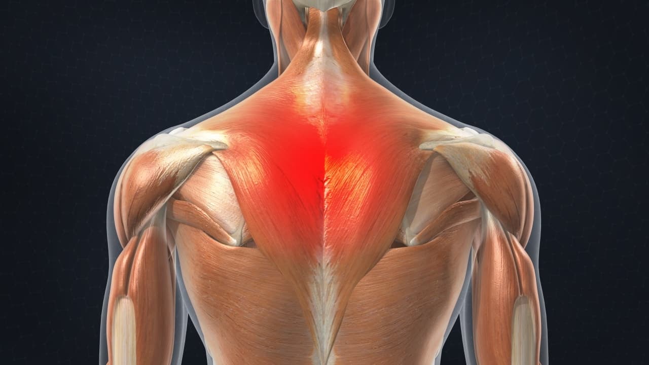
Both the deltoid and the trapezius are firmly attached to the spine of the scapula. Compared to the neck (cervical spine) and lower back (lumbar spine), the upper back is remarkably resistant to injury and pain. The trapezius and latissimus dorsi muscles connect the upper limb to the vertebral column. Back muscles anatomy here include the trapezius, latissimus dorsi, rhomboid and levator scapulae. The muscles of the chest and upper back occupy the thoracic region of the body inferior to the neck and superior to the abdominal region and include the muscles of the shoulders. The traps are quite a complex set of muscles. The muscles of the neck run from the base of the skull to the upper back and work together to bend the head and assist in breathing. The back has different muscle groups that work together to allow movement. Your neck is like no other part of the vertebral spinal. They originate from the vertebrae and insert into the scapulae. The top of the cervical spine connects to the skull, and the bottom connects to the upper back at about shoulder level. The occipital bone surrounds a large opening known as the foramen magnum. 17.11.2015 · neck anatomy explained the neck begins at the base of the skull and connects to the thoracic spine (the upper back).
The majority of these nerves control the functions of the upper extremities and allow you to feel your arms, shoulder, and back of your head. The muscles of the chest and upper back occupy the thoracic region of the body inferior to the neck and superior to the abdominal region and include the muscles of the shoulders. The traps are quite a complex set of muscles. The muscles of the neck run from the base of the skull to the upper back and work together to bend the head and assist in breathing. Neck, in land vertebrates, the portion of the body joining the head to the shoulders and chest.
Pole And Aerial Upper Back Imbalances from static.wixstatic.com (the cervical spine—the neck—has 7 vertebrae, and the lumbar spine—the low back—has 5 vertebrae. The trapezius is one of the broadest and most superficial (closest to the skin) muscles of the upper back and trunk, meaning upon dissection of a cadaver it is often used as a landmark because it is encountered first. These include the anterior, middle and posterior scalene muscles , which extend between the transverse processes of the cervical vertebrae and the upper two ribs. Cervical spine anatomy video the cervical spine has 7 stacked bones called vertebrae, labeled c1 through c7. Upper back pain is also known as thoracic spine pain. The motion of the muscles of the neck are divided into four. The occipital bone is the only bone in your head that connects with your cervical spine (neck). Neck, in land vertebrates, the portion of the body joining the head to the shoulders and chest.
The neck is connected to the upper back through a series of seven vertebral segments.
An area called the occiput. The occipital bone is a bone that covers the back of your head; Each nerve provides sensation to a specific area of the body called a dermatome. The lateral neck muscles, also called the lateral vertebral muscles, are a group of muscles that pass obliquely along the lateral sides of the neck. Cervical spine anatomy video the cervical spine has 7 stacked bones called vertebrae, labeled c1 through c7. It comprises the vertebral column (spine) and two compartments of back muscles; The back is the body region between the neck and the gluteal regions. You have more vertebrae in your thoracic spine than you do in any other spinal region. The cervical spine supports the weight and movement of your head and protects the nerves exiting your brain. Back neck muscles human anatomy course youtube. The trapezius and latissimus dorsi muscles connect the upper limb to the vertebral column. The deltoid, teres major, teres minor, infraspinatus, supraspinatus (not shown) and subscapularis muscles (not shown) all extend from the scapula to the humerus and act on the shoulder joint. The occipital bone is the only bone in your head that connects with your cervical spine (neck).
Anatomy of back of human neck, anatomy of the back and neck, anatomy of the back of the neck, anatomy of the back of the neck muscles, anatomy of the back of your. The top of the cervical spine connects to the skull, and the bottom connects to the upper back at about shoulder level. The lateral neck muscles, also called the lateral vertebral muscles, are a group of muscles that pass obliquely along the lateral sides of the neck. You have more vertebrae in your thoracic spine than you do in any other spinal region. See more ideas about anatomy, shoulder impingement, carpal tunnel.
Take Charge Of Your Upper Back Pain from cloud2.spineuniverse.com Biomechanics and basic anatomy of the lumbar spine / low back. The rhomboid muscle is activated as you bring and squeeze your scapula or shoulder blades back and together. An area called the occiput. You have more vertebrae in your thoracic spine than you do in any other spinal region. The cervical spine protects the nerves connecting to the brain, allowing the head to move freely while supporting its weight. It comprises the vertebral column (spine) and two compartments of back muscles; Priming the pump stretch (back of shoulder). Anatomy of back of human neck, anatomy of the back and neck, anatomy of the back of the neck, anatomy of the back of the neck muscles, anatomy of the back of your.
From the sides and the back of the neck, the splenius capitis inserts onto the head region, and the splenius cervicis extends onto the cervical region.
See more ideas about anatomy, shoulder impingement, carpal tunnel. The muscles of the chest and upper back occupy the thoracic region of the body inferior to the neck and superior to the abdominal region and include the muscles of the shoulders. There's also the sacrum and coccyx, which are 5 fused vertebrae and your tailbone.) the thoracic spine extends from your shoulders to your waist. The occipital bone surrounds a large opening known as the foramen magnum. It also helps extend, tilt, and rotate your neck, which has the effect of bringing your head back, to the side, and turning it. Both the deltoid and the trapezius are firmly attached to the spine of the scapula. It runs from the neck to the upper back. Thoracic vertebrae interlock tightly by overlapping their spinous processes, giving stability to the spine in this region. These muscles give the sides of the neck their. Watch spine anatomy overview video The neck is the area between the skull base and the clavicles. Cervical spine anatomy video the cervical spine has 7 stacked bones called vertebrae, labeled c1 through c7. These include the anterior, middle and posterior scalene muscles , which extend between the transverse processes of the cervical vertebrae and the upper two ribs.
17112015 · neck anatomy explained the neck begins at the base of the skull and connects to the thoracic spine (the upper back) upper back anatomy. The cervical spine protects the nerves connecting to the brain, allowing the head to move freely while supporting its weight.